What is Retinal Vascular Occlusion?
The retina has two main blood vessels. – Central Retinal Artery and Central Retinal Vein. These vessels then form many branches as they travel in the retina.
When the central retina vein is occluded, it is called Central Retinal Vein Occlusion and when the central retinal artery is occluded, it is called Central Retinal Artery Occlusion.
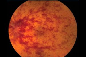
Retinal Vein Occlusion:
When the flow of blood in the retinal vein ( central or its branches) is blocked it is called Retinal Vein Occlusion ( central retinal vein occlusion or branch retinal vein occlusion).
Due to the blockage of the vein, the blood is dispersed over the retina, and in some cases, it is associated with macular edema. The risk factors include diabetes, hypertension, atherosclerosis, inflammatory disorders of blood vessels, dyslipidemia, drugs ( some drugs that are prone to clotting), blood disorders. On diagnosis, a detailed systemic evaluation is done and eye treatment options include intravitreal injection or laser therapy.
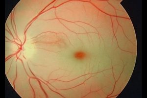
Retinal Artery Occlusion
When the flow of blood in the retinal artery (central or its branches) is blocked, it is called Retinal Artery Occlusion (central retinal artery occlusion or Branch retinal artery occlusion). As the supply is blocked, the area affected gets pale and is deprived of blood. This is an EMERGENCY and needs to be treated urgently to restore the blood flow. The risk factors commonly include dyslipidemia, carotid artery disease, diabetes, hypertension among others. There is a better visual prognosis in patients treated within 6 hours of the incident where the blood reperfusion can be restored.