
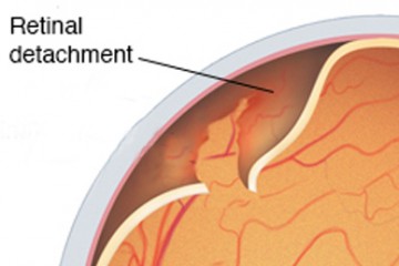
What is Retinal Detachment (RD)?
Retinal detachment is a sight-threatening eye condition and an eye emergency that requires immediate medical attention. This occurs when the retina, the thin, light-sensitive layer at the back of the eye, separates from its underlying tissue. When this delicate tissue pulls away from its normal position and separates from the underlying layers that supply it with oxygen and nutrients, the eye can no longer send proper visual signals to the brain. Once the retina is detached, its cells are cut off from nourishment and start to degenerate quickly, which can lead to permanent vision loss if not treated quickly. This risk of blindness makes early intervention crucial.
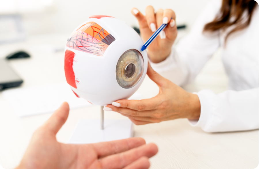
At Vala Eye Centre in Vadodara, Gujarat, our renowned retina specialist, Dr. Ruchi Vala, uses dedicated advanced diagnostic technology and modern safe surgical techniques to repair the retina and preserve your vision. Our superspeciality eye clinic offers comprehensive treatments for a wide range of eye conditions.
Common Causes of Retinal Detachment
Several factors can trigger or increase your risk of developing Retinal Detachment:
- Aging: Natural changes in the gel-like vitreous inside your eye can cause it to shrink and tug on the retina.
- Eye Injury: Trauma or accidents can lead to retinal tears.
- Previous Eye Surgery: Cataract or other intraocular surgeries slightly increase risk.
- Severe Myopia (Nearsightedness): People with high myopia have thinner retinas, making them more prone to tears.
- Diabetic Eye Disease: Proliferative diabetic retinopathy can lead to scar tissue formation and traction on the retina.
- Family History: Genetic factors can also play a role.

Warning Signs You Should Never Ignore
Retinal diseases can affect vision gradually or suddenly, often without noticeable symptoms in the early stages. Retinal detachment symptoms usually develops suddenly and without pain. Early detection is the key to saving your vision. See a retina specialist immediately if you experience any of the following symptoms:
- Sudden appearance or increase in floaters (black spots, cobwebs, or threads in vision.
- Flashes of light, especially in side vision
- A shadow, curtain, or dark patch moving across your vision
- Sudden blurring, distortion of vision and discomfort in the eyes.
These symptoms can appear in one or both eyes. Do not wait for them to go away and timely diagnosis and treatment can make all the difference.
Retinal Detachment can be of three types:
- 1- Rhegmatogenous Retinal Detachment - This type of detachment happens when the vitreous gel in front of the retina starts to liquefy and tries to separate from the retina. However, in some cases, during this separation, it pulls on the retina and creates a break in the retina. The liquified vitreous then start entering the break and goes underneath the retina creating a separation of the retina from its normal position.
- 2 - Tractional Retinal Detachment - The detachment which is triggered by the pull ( traction) from the surface of or over the retina causing it to separate from its normal position is called Tractional RD. This is most commonly seen in people with Vitreous Hemorrhage secondary to Vascular Diseases of the eye like Diabetic Retinopathy.
- 3 - Exudative Retinal Detachment - The detachment where the fluid seeps from under the retina and collects in the subretinal space and separates the retina from its normal position is called Exudative RD. Here, there is no break or traction found. This is generally seen in conditions like Preeclampsia of pregnancy, tumors, uveitic disorders.
The Risk factors include aging eyes ( generally > 50 years), high myopia, family history, trauma over the head or eyes, previous cataracts, or other eye surgeries, in association with other eye diseases like Uveitis, Peripheral Retinal Degeneration.
Diagnosis of Retinal Detachment: What to Expect
A retinal detachment is a sight-threatening emergency that requires a precise clinical diagnosis. Because the symptoms such as blurred vision, sudden onset of floaters or "lightning" flashes can mimic less severe conditions, an immediate specialist evaluation is required to save your vision.
Comprehensive Diagnostic Of Retinal Detachment
- 1- Dilated Fundus Examination: This is the primary diagnostic tool. Your ophthalmologist uses dilating drops to widen the pupil, allowing for an unobstructed view of the eye’s interior. Using indirect ophthalmoscopy, they can scan the entire retinal surface for tears, holes, or fluid accumulation.
- 2- B-Scan Ultrasonography: In cases where a vitreous hemorrhage (blood) or advanced cataracts obscure the doctor's view, ultrasound technology is used. It uses sound waves to create a map of the retina, confirming the detachment even through "cloudy" media.
- 3- Optical Coherence Tomography (OCT): This high-definition, cross-sectional scan allows surgeons to see if the macula (the part of the retina responsible for central vision) is still attached. This "macula-on" or "macula-off" status is the most significant factor in determining the urgency of surgery.
- 4- Scleral Depression: To find small, peripheral tears, the doctor may gently press on the eye's exterior while looking through the lens. This brings the edges of the retina into view.
Retinal Detachment Treatment Options
Dr Ruchi Vala at Vala Eye Centre offers the full range of structured surgical treatments, tailored to the type and severity of the detachment. Each revolutionary method aims and ensures to re-attach the retina and seal any retinal tears.
- 1. Laser Photocoagulation (Retinal Laser) LASIK - Although LASIK Surgery is not the complete solution, surgeons do use specialized medical lasers specifically Argon or Solid-State lasers to address retinal issues through a process called Laser Photocoagulation. This laser treatment is highly effective for sealing retinal tears before they progress into a full detachment.
- 2. Cryotherapy (Freezing Treatment) - Retinal cryopexy is a highly effective clinical technique where specialists apply sub-zero temperatures to stabilize a compromised retina. While refractive surgeries like LASIK focus on the eye's outer lens, cryotherapy reaches the internal posterior layers to facilitate a protective biological bond.
- 3. Pneumatic Retinopexy - Pneumatic retinopexy is a minimally invasive surgical procedure designed to repair specific types of retinal detachment using a specialized gas bubble. Unlike more extensive surgeries like a vitrectomy or scleral buckle, this technique is often performed in an office setting, offering a faster recovery time for eligible patients.
- 4. Scleral Buckling Surgery - Scleral buckling remains a cornerstone surgical intervention designed to repair a rhegmatogenous retinal detachment by physically reducing the internal circumference of the eye. Unlike internal procedures like a vitrectomy, a scleral buckle is an "ab-externo" or external approach that addresses the pathology without necessarily entering the vitreous cavity.
- 5. Vitrectomy Surgery - The primary goal of vitrectomy is to manage "macula-off" scenarios or proliferative vitreoretinopathy, where scar tissue formation complicates traditional repair methods. By clearing the visual path of blood or debris and replacing the natural vitreous with a tamponade agent, surgeons can significantly improve the prognosis for visual recovery.
Why Patients Trust Dr Ruchi Vala for Treating Retinal Detachment in Vadodara
Dr Ruchi Vala has extensive expertise and is trained to diagnose retinal detachments using detailed retinal imaging and to treat them through modern techniques such as vitrectomy, scleral buckle, or pneumatic retinopexy. Our goal is not just to re-attach the retina but also to preserve and restore as much vision as possible through early targeted intervention. Vala Eye Centre is patient centric and dedicated to your eye health and providing exceptional care. We also offer advanced treatments in ophthalmology including contoura for a better lifestyle. While the treatments may involve an injection or be temporarily painful, our team is committed to making the process as comfortable and safe as possible for every patient, including procedures on eyelids.