
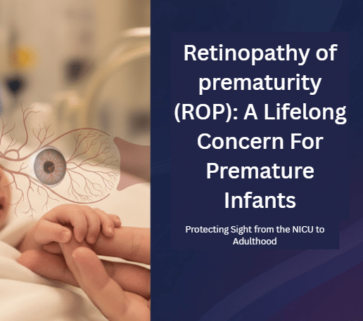
The Neonatal Intensive Care Unit (NICU) is a world defined by paradox. It is a place of profound vulnerability, where life hangs by the thread of a ventilator tube and a premature heartbeat, yet it is also a place of fierce, quiet strength. Here, parents spend hours gazing at their tiny, mighty infants, celebrating every milliliter of milk and every gram of weight gain. In this sacred space of hope and anxiety, there often lurks a silent, insidious threat: Retinopathy of Prematurity, or ROP.
ROP is more than just a medical diagnosis scrawled on a chart; it is a potential fork in the road of a premature infant’s life, carrying implications that extend far beyond the NICU doors and into adulthood. It represents one of the most significant challenges for extremely premature babies—a fight for their sight, the outcome of which shapes their entire existence. While ROP is often successfully treated, transforming what could be a life of darkness into one of sight, it is, fundamentally, a lifelong concern that requires perpetual vigilance, care, and understanding from parents and the medical community alike.
The Science of Interrupted Growth: A Developmental Glitch
To truly appreciate the fight against ROP, we must first understand its origins. It begins with the simple, yet profound, act of premature birth.
In a full-term pregnancy, the retina—the light-sensitive tissue at the back of the eye—doesn’t finish developing its blood vessel network until just before the baby is due. This process starts centrally, near the optic nerve, and grows outwards toward the periphery.
When an infant is born significantly early, this delicate process is abruptly interrupted. Suddenly, the developing retina is exposed to an environment vastly different from the mother’s womb—particularly, a higher level of oxygen.
This change triggers a two-phase, misguided response:
- Phase I: The Vasoconstriction Halt: The higher oxygen levels cause the existing retinal vessels to constrict, or even stop growing entirely, leaving the outer edge of the retina—the peripheral retina—avascular, or without a blood supply.
- Phase II: The Vaso-Proliferative Panic: As the retina matures, it begins to outgrow its current oxygen supply. The avascular peripheral retina, now starved of oxygen, panics and desperately releases excessive amounts of a growth factor known as Vascular Endothelial Growth Factor (VEGF). This sudden flood of VEGF leads to the growth of abnormal, fragile blood vessels—a destructive, uncontrolled sprouting intended to restore blood flow but which instead causes damage.
These abnormal vessels can leak blood and, critically, form scar tissue. As this scar tissue contracts, it pulls on the delicate retina, leading to retinal detachment the most severe outcome and the direct cause of blindness in advanced ROP.
Staging the Disease: From Demarcation Line to Detachment
Ophthalmologists classify ROP by Stages (reflecting severity) and Zones (reflecting location, with Zone I being the most central and critical for good vision).
- Stages 1 & 2 (Mild-to-Moderate): The presence of a faint demarcation line or a raised ridge at the junction of the vascularized and avascular retina. In the vast majority of cases (nearly 90%), ROP resolves spontaneously at these stages without intervention, though ongoing monitoring is essential.
- Stage 3 (Severe): Abnormal fibrovascular tissue begins to grow out of the plane of the retina and into the vitreous humor. This is the point where the disease is considered “treatment-warranted” and intervention is urgently required to prevent retinal detachment. The presence of “Plus Disease” (engorged, twisted vessels indicating significant blood flow issues) pushes the urgency to its absolute maximum.
- Stages 4 & 5 (Advanced): These represent partial or total retinal detachment, often leading to severe vision loss or blindness, even with complex surgical intervention.
The Lifesaving Watch: Timely Screening is Everything
The fight against ROP is a race against time. Because ROP often has no visible external symptoms until it has progressed to severe stages (like a white pupil or unusual eye movements), the only defense is a stringent, timely screening protocol.
This is the job of the dedicated pediatric ophthalmologist, who performs detailed examinations using an indirect ophthalmoscope while the infant is still in the NICU. These exams, though uncomfortable for the baby, are literally sight-saving procedures.
Fresh Update: Evolving Screening Criteria (2024)
Modern medicine constantly refines its understanding of risk. Recent evidence has pushed for a critical re-evaluation of which infants should be screened. While the main criteria remain:
- Gestational Age (GA) less than 31 weeks.
- Birth Weight (BW) less than 1501 grams.
A key 2024 update in some international guidelines, in response to reports of treatable ROP in slightly more mature infants, now includes a recommendation to consider screening infants born between 31+0 and 31+6 weeks’ gestational age.
This shift underscores a crucial point: ROP is a disease that can surprise, and in an era of improving NICU care, the overall health and complex comorbidities of the baby—not just their birth size—must dictate the level of vigilance. Parents must ensure they understand their child’s specific screening schedule, which is based on the baby’s Postmenstrual Age (GA at birth plus postnatal age), a detail often managed by a dedicated ROP coordinator within the hospital.
Treatment: Halting the Progression to Save Central Vision
When ROP progresses to the “treatment-warranted” stage (Type 1 ROP), intervention must be swift, often within 48 to 72 hours. The goal is clear: destroy the avascular, oxygen-starved peripheral retina to stop it from releasing the destructive VEGF, allowing the normal, central part of the retina to survive.
Two primary methods are used today:
- Laser Photocoagulation: This remains the gold standard. A laser is used to strategically burn the avascular peripheral retina. It is a measured sacrifice: some peripheral vision is lost forever, but in doing so, the production of the harmful growth factor stops, halting the disease’s march toward the central retina and preserving the most crucial sight.
- Intravitreal Anti-VEGF Injections: A newer, highly effective approach involves injecting a tiny dose of a drug (an anti-VEGF agent) directly into the eye. This medication acts as a powerful brake, neutralizing the destructive growth factors almost immediately. The advantage is that it can allow for more normal, slow regrowth of vessels, potentially minimizing the loss of peripheral vision. However, the long-term safety and systemic effects of these powerful drugs on a tiny, developing infant are still under extensive study, making it a treatment option that requires careful consideration.
In the more advanced, rare stages (Stage 4 and 5), complex retinal surgery, such as a vitrectomy (removing scar tissue) or a scleral buckle (a band placed around the eye to ease traction), may be necessary, though the prognosis for functional vision is significantly lower at these late stages.
ROP: A Lifelong Concern for the Former Preemie
The NICU stay is a chapter, but for infants who had ROP, the story continues. The “all clear” upon discharge does not mean the end of their visual journey. ROP, even in its mildest forms that resolve spontaneously, is a permanent marker on a child’s retina, and it drastically increases the risk for a host of secondary eye problems that manifest months, years, or even decades later.
This is the meaning of the “lifelong concern.”
The Common Long-Term Complications
- Myopia (Nearsightedness): This is the single most common long-term complication, occurring in a significant percentage of former ROP patients, often severely. The healing and scarring process of the retina changes the shape of the eye, making it focus images in front of the retina rather than on it. Regular vision checks are non-negotiable.
- Strabismus (Misaligned Eyes) and Amblyopia (Lazy Eye): The scarring and pulling on the retina can also lead to the eyes being misaligned (strabismus) and, consequently, the brain beginning to ignore the input from the weaker eye (amblyopia). Treatment often involves glasses, patching, or eye muscle surgery.
- Glaucoma and Cataracts: The complex history of inflammation and intervention in the eye can increase the risk for both glaucoma (damage to the optic nerve from high eye pressure) and cataracts (clouding of the lens) later in life.
- Late Retinal Detachment: The peripheral retina in ROP survivors, even those successfully treated, is often thinner and more prone to tears and holes. An area of avascular retina or a band of scar tissue creates a weak point. Years later, a minor injury or just the natural aging of the vitreous gel can pull on this weak spot, causing a new, devastating retinal detachment. This risk persists into adulthood.
Beyond the Eye: The Neurodevelopmental Connection
Adding to the complexity, research continues to explore the intricate connection between severe ROP and broader neurodevelopmental outcomes. While some studies suggest severe ROP is not an independent predictor of worse cognitive or motor scores when other factors of extreme prematurity are controlled for, it is an undeniable marker of a fragile infancy. The same underlying physiological stressors—oxidative stress, inflammation, and fluctuations in oxygen levels—that disrupt retinal growth are also implicated in brain development. Therefore, a diagnosis of severe ROP is often a flag for the need for comprehensive, multidisciplinary follow-up covering cognitive, motor, and behavioral development.
The Power of Vigilance and Community
The story of ROP is ultimately a story of human resilience—the incredible spirit of the smallest babies and the unwavering dedication of the medical teams who fight for them.
For the parents, the journey demands extraordinary commitment: the trauma of the NICU is followed by a life of routine, specialized eye exams. It means trusting a pediatric ophthalmologist who becomes a lifelong partner in your child’s care, conducting annual check-ups well into adulthood to screen for the secondary complications that are an inevitable risk.
The medical community, in turn, must champion comprehensive, multidisciplinary follow-up. It is a commitment that extends from the neonatologist who manages oxygen to the ROP coordinator who schedules the crucial first exam, and finally to the adult ophthalmologist who understands the unique risks of the former premature retina.
The fight against blindness from ROP is largely won in the NICU, but the care for the ROP survivor is a marathon, not a sprint. By understanding that Retinopathy of Prematurity is not just an infant disease, but a lifelong condition that requires sustained awareness, we can ensure that these tiny fighters—our beloved premature infants—grow up not only seeing but thriving, with their futures as bright as their resilience is strong.