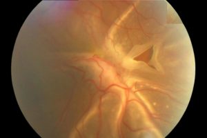
What is Retinal Detachment (RD)?
Retinal detachment is a sight-threatening eye condition and an eye emergency that requires immediate medical attention. This occurs when the retina, the thin, light-sensitive layer at the back of the eye, separates from its underlying tissue. When this delicate tissue pulls away from its normal position and separates from the underlying layers that supply it with oxygen and nutrients, the eye can no longer send proper visual signals to the brain. Once the retina is detached, its cells are cut off from nourishment and start to degenerate quickly, which can lead to permanent vision loss if not treated quickly. This risk of blindness makes early intervention crucial.
At Vala Eye Centre in Vadodara, Gujarat, our renowned retina specialist, Dr. Ruchi Vala, uses dedicated advanced diagnostic technology and modern safe surgical techniques to repair the retina and preserve your vision. Our superspeciality eye clinic offers comprehensive treatments for a wide range of eye conditions.
Common Causes of Retinal Detachment
Several factors can trigger or increase your risk of developing RD:
-
Aging: Natural changes in the gel-like vitreous inside your eye can cause it to shrink and tug on the retina.
-
Eye Injury: Trauma or accidents can lead to retinal tears.
-
Previous Eye Surgery: Cataract or other intraocular surgeries slightly increase risk.
-
Severe Myopia (Nearsightedness): People with high myopia have thinner retinas, making them more prone to tears.
-
Diabetic Eye Disease: Proliferative diabetic retinopathy can lead to scar tissue formation and traction on the retina.
-
Family History: Genetic factors can also play a role.
Warning Signs You Should Never Ignore
Retinal detachment symptoms usually develops suddenly and without pain. Early detection is the key to saving your vision.
See a retina specialist immediately if you experience any of the following symptoms:
-
Sudden appearance or increase in floaters (black spots, cobwebs, or threads in vision)
-
Flashes of light, especially in side vision
-
A shadow, curtain, or dark patch moving across your vision
-
Sudden blurring or distortion of vision
These symptoms can appear in one or both eyes. Do not wait for them to go away and timely diagnosis and treatment can make all the difference.
Diagnosis: How We Detect this condition
At our retina clinic in Vadodara, we use advanced diagnostic tools to confirm and evaluate retinal detachment:
-
Dilated Retinal Examination: Using special lenses and bright light to view the retina in detail.
-
Optical Coherence Tomography (OCT): A non-invasive scan that shows high-resolution cross-sections of the retina.
-
B-Scan Ultrasonography: Helpful when the view is blocked by bleeding or cataract.
Early diagnosis allows for simpler and more effective treatment, often preventing total vision loss.
Retinal Detachment Treatment Options
Our retina specialists offer the full range of surgical treatments, tailored to the type and severity of the detachment. Each method aims to reattach the retina and seal any retinal tears.
1. Laser Photocoagulation (Retinal Laser) LASIK
Used for small retinal tears or holes before detachment progresses. A focused laser creates tiny burns around the tear, forming a scar that seals the retina to the back of the eye. This is an outpatient procedure with minimal discomfort and quick recovery.
2. Cryotherapy (Freezing Treatment)
An alternative to laser for certain retinal breaks. A freezing probe is used to create a scar that “glues” the retina back in place. This method is often combined with other surgical techniques.
3. Pneumatic Retinopexy
A minimally invasive procedure performed under local anesthesia. The surgeon injects a small gas bubble into the eye, which gently pushes the retina against the wall. The tear is then sealed with laser or cryotherapy. Patients must maintain a specific head position for several days to allow proper healing.
4. Scleral Buckling Surgery
In this time-tested procedure, a flexible silicone band (buckle) is placed around the white of the eye (sclera). It indents the eye wall slightly, reducing traction and helping the retina settle back into place. This is often recommended for younger patients or complex tears.
5. Vitrectomy Surgery
This is the most common and effective treatment for larger or complex retinal detachments. The surgeon removes the vitreous gel, replaces it with a clear solution or gas, and reattaches the retina using laser or cryotherapy. Vitrectomy offers precise control and excellent long-term results when performed by experienced retina surgeons.
Retinal Detachment can be of three types:
- Rhegmatogenous RD: This type of detachment happens when the vitreous gel in front of the retina starts to liquefy and tries to separate from the retina. However, in some cases, during this separation, it pulls on the retina and creates a break in the retina. The liquified vitreous then start entering the break and goes underneath the retina creating a separation of the retina from its normal position.
- Tractional RD: The detachment which is triggered by the pull ( traction) from the surface of or over the retina causing it to separate from its normal position is called Tractional RD. This is most commonly seen in people with Vitreous Hemorrhage secondary to Vascular Diseases of the eye like Diabetic Retinopathy.
- Exudative RD: The detachment where the fluid seeps from under the retina and collects in the subretinal space and separates the retina from its normal position is called Exudative RD. Here, there is no break or traction found. This is generally seen in conditions like Preeclampsia of pregnancy, tumors, uveitic disorders.
The Risk factors include aging eyes ( generally > 50 years), high myopia, family history, trauma over the head or eyes, previous cataracts, or other eye surgeries, in association with other eye diseases like Uveitis, Peripheral Retinal Degeneration.
The Role of Our Retina Specialists
At Vala Eye Centre, an advanced retina clinic in Vadodara Gujarat, our vitreoretinal specialists Dr Ruchi Vala have extensive expertise and are trained to diagnose retinal detachments using detailed retinal imaging and to treat them through modern techniques such as vitrectomy, scleral buckle, or pneumatic retinopexy. Our goal is not just to reattach the retina but also to preserve and restore as much vision as possible through early, targeted intervention. We are dedicated to your eye health and providing exceptional care. We also offer advanced treatments in ophthalmology including contoura for a better lifestyle. While the treatments may involve an injection or be temporarily painful, our team is committed to making the process as comfortable and safe as possible for every patient, including procedures on eyelids.